
.jpeg)
How to detect liver cancer?
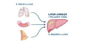
Liver cancer is a malignant tumor of the liver. Early detection and accurate diagnosis are crucial to improving the survival rate of patients. With the development of medical technology, the detection methods for liver cancer have become increasingly diversified. Detection of liver cancer usually involves a variety of methods, and different examinations can provide different information to help doctors determine the severity of the disease, treatment options, and prognosis. Common examination methods mainly include blood tests, ultrasound, CT scans, MRI, biopsies, etc.
When diagnosing liver cancer, the first thing done is a blood test. This test evaluates liver function, looks at blood clotting, and detects the presence of certain biomarkers, such as alpha-fetoprotein (AFP), a tumor marker associated with liver cancer. Blood tests can also be used to screen for hepatitis B or hepatitis C virus infections, which are important causes of liver cancer. If blood tests show abnormalities, your doctor may recommend further imaging tests.
Ultrasound examination is one of the most common methods for diagnosing primary liver cancer. By using high-frequency sound waves, ultrasound can produce images of the inside of the liver, helping doctors see the size, shape and presence of abnormal tissue. This method is non-invasive and can be completed quickly, allowing for easy initial screening.
If the ultrasound results are suspicious, a CT scan may be performed as a next step. CT scans can produce three-dimensional images of multiple structures surrounding the liver, helping doctors determine whether the cancer has spread to other organs. Compared with ultrasound, CT scans have higher resolution and can provide more detailed information, which is helpful in formulating surgical plans.
In addition, magnetic resonance imaging (MRI) is also an important method for detecting liver cancer. MRI scans use strong magnetic fields and radio waves to produce detailed images of the inside of the body, which can clearly show the extent of the tumor and its impact on surrounding major blood vessels. In some cases, PET-CT scans are also used to check for the presence of secondary liver cancer, helping doctors understand the systemic condition of the cancer.
Biopsy is an important way to diagnose liver cancer. By removing liver tissue samples and examining them under a microscope, it can be determined whether there are cancer cells. This examination usually uses either fine needle aspiration or laparoscopic surgery. Fine-needle aspiration uses a fine needle to extract liver cells under local **, while laparoscopic surgery uses a small incision to directly observe the liver and obtain tissue samples under general anesthesia.
Finally, if a patient is diagnosed with secondary liver cancer, additional tests may be needed to determine the source of the primary cancer. These examinations will help doctors develop individualized treatment plans to improve patients' quality of life and survival rates.
In short, the diagnosis process of liver cancer is complex and professional, and requires a comprehensive evaluation using a variety of examination methods. After diagnosis, patients may experience a period of mood swings. It is recommended to actively communicate with doctors and family members to face this challenge together and find a suitable treatment plan.
(Click to view an introduction to drugs for treating liver cancer.)
xa0
References:
https://www.cancer.org.au/cancer-information/types-of-cancer/liver-cancer
[ 免责声明 ] 本页面内容来自公开渠道(如FDA官网、Drugs官网、原研药厂官网等),仅供持有医疗专业资质的人员用于医学药学研究参考,不构成任何治疗建议或药品推荐。所涉药品可能未在中国大陆获批上市,不适用于中国境内销售和使用。如需治疗,请咨询正规医疗机构。本站不提供药品销售或代购服务。


