
.jpeg)
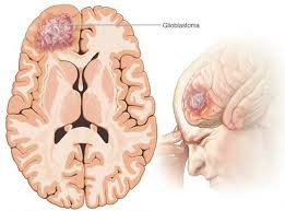
Imaging manifestations of glioblastoma (GBM)
Glioblastoma (GBM) is the most malignant central nervous system tumor, and its imaging manifestations are an important basis for diagnosis and treatment decisions. Commonly used imaging tests include magnetic resonance imaging (MRI), computed tomography (CT), and positron emission tomography (PET). The following is an analysis of the specific performance and characteristics of each imaging method:
1. Magnetic resonance imaging (MRI)
MRIis the preferred imaging method for diagnosing GBM because of its high-resolution and multi-parameter imaging capabilities, which can comprehensively display the shape, boundaries and invasion of surrounding tissues of the tumor.
1.1 Plain scan (T1 and T2 weighted imaging)
T1weighted imaging (T1WI): The parenchymal portion of the tumor usually appears as a low-signal area, while areas of hemorrhage or necrosis may appear as high-signal.
T2weighted imaging (T2WI): Tumors and surrounding edema areas mostly show high signal, which is in obvious contrast with normal brain tissue. Peripheral edema is widespread and is often caused by tumor infiltration and increased vascular permeability.
1.2 Enhanced scanning
The typical manifestation of GBM is that the tumor shows annular or irregular morphological enhancement, with no enhancement in the central necrotic area and obvious edge enhancement, indicating that the blood-brain barrier is destroyed. Extensive T2high-intensity edema areas are common outside the enhancement area, reflecting the infiltrative growth characteristics of tumors into surrounding tissues.
1.3 Functional Imaging
Diffusion-weighted imaging (DWI): Central necrotic areas usually show high diffusion signal (ADC values u200bu200bincrease), while areas with higher tumor cell density show restricted diffusion (ADC values u200bu200bdecrease).
Perfusion-weighted imaging (PWI): GBM usually manifests as a significant increase in relative cerebral blood volume (rCBV), reflecting tumor neovascularization.
Magnetic resonance spectroscopy (MRS): The MRS signature of GBM includes choline (Cho) peak increased, N-acetyl aspartate (NAA) peak decreased, and lactic acid and lipid peaks appeared, indicating active tumor metabolism and necrosis.
2. Computed tomography (CT)
CT is often used for initial evaluation in the emergency setting, especially if increased intracranial pressure, cerebral hemorrhage, or bone destruction is suspected. GBM’s typical CT manifestations include:
Low-density lesions: Tumor necrosis areas are mostly low-density areas with extensive surrounding edema areas.
Ring enhancement: During enhanced scan, irregular ring enhancement of the tumor can be seen, and there is no enhancement in the central necrotic area.
Hemorrhage or calcification: SomeGBM patients may have punctate or flake-like high-density areas in their tumors, indicating intratumoral hemorrhage or calcification.
AlthoughCTis not as good asMRI in distinguishing tumor details, it has the advantages of speed and convenience in emergency patients.
3. Positron emission tomography (PET)
PETimaging reflects the metabolic activity of tumors through tracers (such as 18F-FDG), which can help evaluate the malignancy of GBM and identify recurrence and changes after treatment.
18F-FDG PET: GBM usually exhibits high metabolic activity, with significantly increased uptake in tumor areas, in sharp contrast to normal brain tissue.
Amino acidsPET (such as 11C-MET or 18F-FET)< /span>: Amino acidsPET is more sensitive to the diagnosis of GBM and can display tumor boundaries and low metabolism areas, especially when evaluating recurrence.
4. Typical features of imaging findings
GBMThe imaging features of GBM are complex due to their high heterogeneity, but the following points are typical manifestations:
Ring enhancement: The central necrotic area and enhanced edge reflect the characteristics of tumor angiogenesis and necrosis.
Invasive growth: Tumors often spread through white matter fiber tracts, resulting in unclear boundaries on images.
Peripheral edema: The T2 high signal area around the tumor indicates edema caused by tumor infiltration or increased vascular permeability.
Multifocal lesions: Multiple lesions may be seen in some patients, suggesting possible spread through cerebrospinal fluid.
5. Application of imaging in treatment
Imaging tests are not only used for diagnosisGBM, also plays an important role in treatment planning and efficacy evaluation:
Preoperative evaluation:MRI can accurately display tumor boundaries and important functional areas, providing reference for surgical planning.
Radiation therapy planning: Imaging helps accurately outline the target area and optimize the radiotherapy plan.
Response Monitoring:MRIenhanced scanning or PET imaging can be used to assess residual tumor or recurrence after treatment.
The imaging manifestations of glioblastoma are complex and highly heterogeneous, but MRI enhanced scanning and functional imaging can more comprehensively display tumor characteristics and are core tools for diagnosis and treatment. Combining CT and PET can more accurately assess the distribution, metabolism and invasiveness of tumors, providing an important basis for personalized treatment.
(Click to view an introduction to drugs for the treatment of glioblastoma.)
xa0
Reference materials
1.National Cancer Institute. Imaging Techniques in Glioblastoma. https://www.cancer.gov
2.American Brain Tumor Association. Diagnostic Imaging for GBM. https://www.abta.org
3.UpToDate. Imaging Features of Glioblastoma. https://www.uptodate.com
[ 免责声明 ] 本页面内容来自公开渠道(如FDA官网、Drugs官网、原研药厂官网等),仅供持有医疗专业资质的人员用于医学药学研究参考,不构成任何治疗建议或药品推荐。所涉药品可能未在中国大陆获批上市,不适用于中国境内销售和使用。如需治疗,请咨询正规医疗机构。本站不提供药品销售或代购服务。


