
.jpeg)
How to detect endometrial cancer
Early detection of endometrial carcinoma (endometrial carcinoma) is crucial to improve treatment effectiveness and prognosis. The disease is usually diagnosed through a series of tests and evaluations. First, patients often present with symptoms such as irregular vaginal bleeding, prolonged menstruation, or excessive vaginal discharge. These symptoms may indicate abnormal changes in the endometrium, prompting patients to seek medical help.
During the initial evaluation, the doctor will conduct a detailed history and physical examination. The medical history includes the patient's menstrual cycle, childbirth history, past diseases and family medical history. Physical examination is mainly to observe the status of the vulva, vagina or cervix through gynecological examination. During this stage, doctors may notice some suspicious signs, but they won't be able to diagnose it.
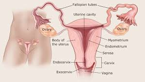
To further confirm the diagnosis, doctors often recommend an ultrasound, specifically a transvaginal ultrasound (TVUS). This examination can clearly show the thickness of the endometrium. If the thickness of the endometrium is significantly thicker (usually greater than 4-5 mm) or there are other abnormal changes, this may be a sign of endometrial cancer. Although ultrasound examination is a non-invasive detection method, it cannot confirm the diagnosis and can only be used as the basis for subsequent examinations.
In cases where ultrasound results are suspicious, doctors often recommend an endometrial biopsy. This is the gold standard for diagnosing endometrial cancer, by which endometrial tissue can be obtained for pathological examination. A biopsy is usually done in an outpatient setting, where the doctor uses a fine needle or a dilation and curettage device to take a sample. The tissue sample obtained is analyzed under a microscope to determine the presence and type of malignant cells.
In addition to the above examinations, in some cases the doctor may recommend a hysteroscopy, which is a more direct examination method. Through hysteroscopy, doctors can directly observe the state of the endometrium and perform a biopsy at the same time. Hysteroscopy can provide a clearer view and help detect local lesions.
In addition, depending on the patient's specific situation, the doctor may also perform imaging tests, such asCT scan or MRI, to evaluate the spread of the tumor. This is of great significance in determining the stage of the disease and treatment options.
(Click to view an introduction to drugs for the treatment of endometrial cancer)
xa0
References: https://www.cancer.org/cancer/types/endometrial-cancer/detection-diagnosis-staging/how-diagnosed.html
[ 免责声明 ] 本页面内容来自公开渠道(如FDA官网、Drugs官网、原研药厂官网等),仅供持有医疗专业资质的人员用于医学药学研究参考,不构成任何治疗建议或药品推荐。所涉药品可能未在中国大陆获批上市,不适用于中国境内销售和使用。如需治疗,请咨询正规医疗机构。本站不提供药品销售或代购服务。


